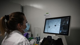
Researchers from the University of Adelaide have developed a high-tech medical device in an effort to increase safety during brain surgery.
Called a "smart needle," the device is a small imaging probe. Fitted inside a brain biopsy needle, it provides surgeons a look at at-risk blood vessels when the needle is inserted, helping prevent potentially fatal bleeding in the brain. Aside from offering surgeons a view of the surgical site, the device is also able to recognize blood vessels on its own, giving out warnings when at-risk blood vessels are in proximity.
Robert McLaughlin, Biophotonics chair at the University of Adelaide's Center for Nanoscale Biophotonics, explained that the smart needle contains a small fiber optic camera that's just the size of a human hair. It works by shining infrared light on the intended area to let surgeons see the blood vessels before they are accidentally damaged.
The smart needle was used over the course of the last six months on 12 patients for a pilot trial program at Western Australia's Sir Charles Gairdner Hospital.
Developing The Smart Needle
The smart needle and the laboratory it was developed in was shown to Australia Sen. Simon Birmingham on Jan. 20. The lab received partial funding support from the South Australian Government, the National Health and Medical Research Council, and the Australian Research Council.
According to Birmingham, the Turnbull Government had entered into a $23-million commitment to spur vital research until 2021 via the Australian Research Council's Center of Excellence for Nanoscale BioPhotonics. He also cited the smart needle as a perfect representation of how research investments can lead to real benefits and improve the lives of Australians.
"We will see this as one of the first in the next generation of research breakthroughs supported by the Turnbull Government's National Innovation and Science Agenda," he added.
The smart needle is set to begin formal clinical trials in 2018 and discussions are in place to manufacture the medical device in Australia.
Watch McLaughlin discuss their work on the smart needle below!
Other Advancements In Brain Surgery
Last year in December, neurosurgeons from Cedars-Sinai Medical Center unveiled a high-definition imaging device called the BrightMatter Guide that will allow them to see inside a human brain during surgery. With it, surgeons will be able to map pathways safely, offering easier access for reaching and removing tumors.
In the United States alone, about 62,000 primary tumors and 150,000 metastatic tumors in the brain are diagnosed every year. With these many potential brain surgeries, the BrightMatter Guide offers hope for better outcomes.
Cedars-Sinai is the first facility to use the 3D imaging technology in California to carry out brain surgeries. Seeing some 600 brain surgeries each year, the hospital's medical staff will now be able to assess in real-time any issues that may occur during the procedure.
Before the BrightMatter Guide, surgeons had trouble creating MRI mapping techniques that would be able to scan brain tumors in better detail. The new 3D imaging device may also be considered as a replacement for surgical microscopes as it offers a more sensitive means of scanning the brain, which can affect how the surgical procedure turns out.


