Scientists have tracked the reaction of a protein in response to light, leading us to further understand essential, life-sustaining processes including human vision.
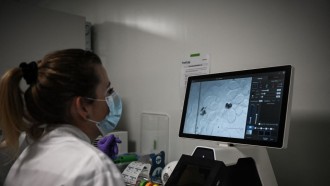
An international team of researchers led by the University of Wisconsin-Milwaukee and Imperial College London captured images of a tiny crystallized protein — or 25 trillion per second — as it reacted to light.
Deemed the first instantaneous look at the mechanisms driving a chemical reaction, the experiment used the world’s most potent X-ray laser at the SLAC National Accelerator Laboratory and allowed them to witness atomic movements as fast as 100 femtoseconds or quadrillionths of a second — a thousand times faster than ever before. The snapshots were taken in a bacterial light sensor.
“This puts us dramatically closer to understanding the chemistry necessary for all life,” said study author and physics professor Marius Schmidt. “Discovering the step-by-step process of how proteins function is necessary not only to inform treatment of disease, but also to shed light on the grand questions of biology.”
According to co-author Dr. Jasper van Thor, the protein structure can usually only be imaged post-reaction. Their work is deemed the first time that crystal structures are imaged on timescales where the proteins are still going through the reaction — from theory and spectroscopy to actual reality, he added.
The novel technique could benefit a wide range of light-induced, ultra-fast atom-level motions. For instance, it could show how the human eye’s visual pigments react to light along with how too much absorption of it leads to their damage. It could also reveal how atomic structures react to pulses of light with varying shapes and duration, a crucial first step in manipulating chemical reactions using light.
The data demonstrated how the bacterial sensor reacts right away after light absorption. The team observed them by focusing on the light-sensitive section of the protein dubbed “photoactive yellow protein” (PYP), which works as an eye in purple bacteria. This helps the bacteria “feel” blue light and avoid excessively energetic, likely harmful light.
Using the Linac Coherent Light Source X-ray (LCLS) Free Electron Laser situated in California, the group fired extremely bright pulses at the protein, capturing images every few femtoseconds during the photon reaction’s progression.
X-ray pulses of the LCLS are very short and last a mere couple of femtoseconds, thus allowing the team to investigate processes on that timescale. Tweaking an earlier experiment, the scientists replaced optical laser with a new one boasting of being 100,000 times shorter than earlier and better approximates the length of the X-ray pulse.
They applied more efficient timing tools as well in order to measure the relative time of arrival between the optical and X-ray laser pulses. This enhances the capacity to track mega-fast events.
The findings were published May 5 in the journal Science.