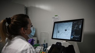
Studying how nerve cells in the brain communicate with each other by transmitting chemical signals through synapses can be very difficult and much so when the process, known as synaptic transmission, is observed in living animals.
A group of researchers, however, reported that they have successfully observed this particular neuron activity with the help of a technique that uses genetics and the physics of light.
Aurélie Pala and Carl C.H. Petersen from the Ecole Polytechnique Federale de Lausanne (EPFL) in Switzerland reported that they were able to observe the chemical signals that nerve cells send to one another through the help of a technique dubbed optogenetics.
The process involves inserting a gene of a light-sensitive protein into living nerve cells. The light-sensitive protein eventually sits on the cell membrane, where it functions as an electrical gate that opens up and permits electrical ions to enter into the cells when light is shone upon it.
The electrical flow alters the voltage balance of the neurons, allowing the researchers to control the activity of certain neurons in living and moving animals. This is crucial in studying different types of neurons and understanding brain functions such as memory, thought, language and behavior.
For their study published in the journal Cell on Dec. 24, Pala and Petersen used optogenetics to target the neurons of experimental rodents. The particular neurons are found in the region of the brain known as barrel cortex, which is responsible for handling stimuli from the animals' whiskers.
The genetically modified neurons produced electrical signals when the researchers shone a blue light into them, allowing the researchers to look at the synaptic transmission from the light sensitive nerve cells to small connector neurons known as interneurons. By using an advanced imaging technique, the researchers were also able to identify each of the interneurons.
"This is a proof-of-concept study," Pala said. "Nonetheless, we think that we can use optogenetics to put together a larger picture of connectivity between other types of neurons in other areas of the brain."
The study involved anesthetized mice, but the researchers said they were planning to try the technique on non-sedated mice in order to see how neuronal switch influences higher brain functions.
"We therefore demonstrate the technical feasibility of assessing functional cell-type-specific synaptic connectivity in vivo, allowing future investigations into context-dependent modulation of synaptic transmission," the researchers wrote.