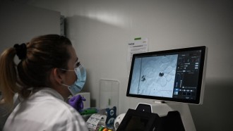
After decades of extensive research, scientists have finally discovered a way to observe neural electricity in an actual living creature.
Adam Cohen, professor of chemistry and chemical biology and of physics at Harvard, first author Dr. Yoav Adam, and their cross-disciplinary research team have managed to transform neural electrical signals into sparks visible through a microscope by shedding light on the brain.
The research was published in the scientific journal Nature on Wednesday, May 1.
Busy Brain
According to scientists, observing a real live session of neural electricity is just like watching a live broadcast of the brain. Since neurons are responsible for every thought and sensation living creatures feel, they send and receive massive amounts of information, which is still mostly incomprehensible to scientists until recently.
Electrical signals can travel from cell to cell at up to 270 miles per hour. At that kind of speed, trying to see neural electricity inside a busy brain is just like trying to see the electricity inside a telephone wire, which no naked eye can achieve.
So for scientists to observe firsthand how neurons turn information into behaviors, emotions, and thoughts, they created a particular procedure for them to see.
Talented Protein
Cohen got the inspiration from another study made by the researchers at the Massachusetts Institute of Technology in 2010. In the study, the MIT researchers introduced the protein Archaerhodopsin 3 to a brain and caused it to light up using a special tool. The tool then converted the light into electricity.
Archaerhodopsin 3 and its host organism was discovered by an Israeli ecologist in an ecological survey in 1980s. The organism is able to convert sunlight into electrical energy in a primitive form of photosynthesis because of the protein.
After years of studying, Cohen and his team found a way to reverse the organism's trick and use the protein to convert the electrical activity of neurons into observable flashes of light.
Red And Blue Light
Cohen and his team were able to manipulate Archaerhodopsin 3 to turn voltage into light when illuminated by a red light. According to the study, in that way, the protein will act as an ultra-sensitive voltmeter that changes with an electric jolt.
The team then paired Archaerhodopsin 3 with a similar protein that sparks electrical impulses in the neurons when illuminated with blue light. According to Adam, that particular process is vital for recording and controlling the cells' activities.
The red light is responsible for recording, and the blue light is responsible for controlling. Although the paired proteins work well in a dish, it was a real challenge for Cohen and his team to make the process work inside a living brain.
Making A Little Movie
It was five years of intensive research and interdisciplinary collaboration between statisticians, physicists, biochemists, computer scientists, molecular biologists, and 24 neuroscientists before the whole team managed to perform the experiment successfully in the brain of a living mouse.
By tweaking the proteins to work in a mouse brain, positioning the proteins carefully with genetic manipulation, and making a new microscope with a video projector specific for the whole process, they were able to glean positive results.
"You basically make a little movie," says Cohen.
The study is just the first step of many, according to Cohen and his team.
"A mouse brain has 75 million cells in it. So depending on your perspective, we've either done a lot or we still have quite a long way to go," added Cohen. The rest of the team is working on improving their software and tools to record the process clearer and on a broader scope.
According to Adam, he's positive that further study could help them reach maximum results.


