Scientists from UC Davis have utilized dynamic total-body positron emission tomography (PET) to capture the immune response of the human body to COVID-19 infection in recovering patients.
The study offers a pioneering look into how the immune system reacts to viral infections and establishes long-term protection against re-infection.
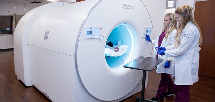
Novel Imaging Tech
The researchers employed the uEXPLORER total-body PET scanner, a cutting-edge imaging technology developed in collaboration with United Imaging Healthcare at UC Davis.
This technology involves the injection of minute amounts of radiotracers into the patient, followed by continuous imaging over a period, essentially creating a 'movie' of the radiotracer's distribution over time within the body.
This data is then used for biologically-relevant analysis through mathematical models.
Total-body PET scanners facilitate simultaneous dynamic imaging and kinetic modeling in all body organs. They possess significantly greater sensitivity compared to conventional PET systems, resulting in superior image quality while allowing the use of lower radiotracer doses, according to the team.
Negar Omidvari, assistant project scientist in the UC Davis Department of Biomedical Engineering, emphasized that dynamic total-body PET is currently the only technology with an acceptable radiation dose that enables noninvasive quantitative measurements of immune cell distribution and movement within all tissues in living humans.
This study marks the first application of dynamic PET and kinetic modeling to measure CD8+ T cell distribution in humans. These specific immune cells, characterized by the CD8 protein on their surface, play a crucial role in identifying and eliminating infected cells during a viral infection.
Some of these CD8+ cells transform into antigen-specific memory T cells, providing long-term protection against re-infection.
For the study, three healthy individuals and five recovering COVID-19 patients with mild or moderate symptoms were enrolled. Participants were injected with a small quantity of a radioactive liquid containing an immunoPET radiotracer targeting human CD8. A series of dynamic scans were then conducted over specified time intervals.
The researchers meticulously assessed radiotracer activity in both blood and non-blood tissue on PET images. Kinetic modeling was used to differentiate the impact of blood circulation on tissue, enabling measurement of radiotracer uptake in tissues irrespective of imaging timing or individual participant blood variations.
The results demonstrated high CD8+ T cell uptake in the lymphoid organs of all participants, with the highest in the spleen, followed by the bone marrow, liver, tonsils, and lymph nodes.
Notably, recovering COVID-19 patients exhibited increased CD8+ T cell concentrations in the bone marrow compared to healthy controls. In follow-up imaging at 6 months post-infection, these concentrations were slightly higher than those at around 2 months post-infection.
Promising Platform
This research presents a promising platform for non-invasive, long-term assessment of T cell distribution across the entire human body, showcasing its potential in studying immune responses comprehensively.
The study's findings hold implications for investigating immune memory, treatment response in cancer patients, infectious diseases, autoimmune disorders, transplant procedures, and therapeutic and vaccine development.
The study's findings were published in Science Advances.
Related Article : Eating Disorders Among Female Adolescents Increased During the COVID-19 Pandemic, New Research Finds

ⓒ 2025 TECHTIMES.com All rights reserved. Do not reproduce without permission.


