Scientists have achieved a breakthrough in medical imaging, producing the sharpest-ever scan of a brain.
The scan, which is 64 million times clearer than a typical clinical MRI, was created by a team from Duke's Center for In Vivo Microscopy, working in collaboration with researchers from the University of Tennessee Health Science Center, the University of Pennsylvania, University of Pittsburgh, and Indiana University.
Incredibly Powerful Magnet
The researchers used an incredibly powerful magnet, 100 times stronger than those used in clinical MRI machines, and a high-performance computer that was the equivalent of 800 laptops working together to produce the scan.
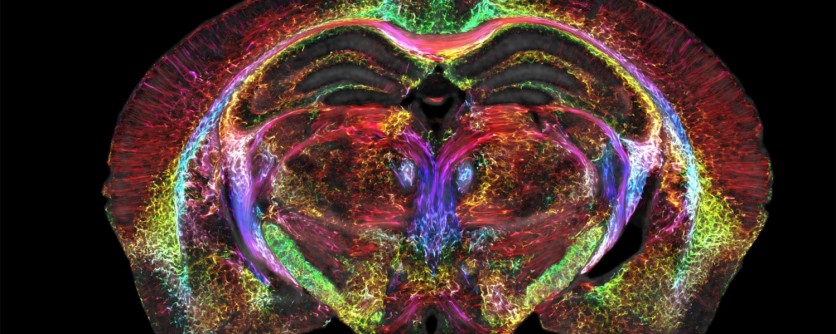
The images are so detailed that they can reveal microscopic details within the brain, offering a new way to visualize the connectivity of the entire brain at record-breaking resolution.
The images were created by the researchers using mice rather than people. Still, the discovery will provide a better knowledge of human conditions, such as how the brain changes with aging, a person's diet, or neurodegenerative disorders like Alzheimer's.
The development of the high-resolution MRI has taken nearly 40 years, with the team at the Duke Center for In Vivo Microscopy perfecting numerous components.
To photograph one brain, the researchers used an extraordinarily strong magnet, a unique combination of gradient coils that are 100 times more powerful than those used in a clinical MRI, and a high-performance computer equivalent to approximately 800 laptops.
Major Implications of These Scans
The latest scans have major implications for research into human disorders like Huntington's, Alzheimer's, and others. It is hoped that the increased power of the microscope will enhance the comprehension of how comparable mechanisms operate or malfunction in humans.
One set of MRI images demonstrates how the connectivity of the mouse brain changes over time, while another set demonstrates how specific regions, such as the subiculum, which is important in memory, change more than the rest of the mouse brain.
Researchers were able to display a spool of rainbow-colored brain connections in a mouse model of Alzheimer's disease, emphasizing the astonishing decline of neural networks.
The discovery presents fresh opportunities for the medical community to investigate and comprehend neurological diseases in unprecedented depth and open the door for novel therapies.
"It is something that is truly enabling. We can start looking at neurodegenerative diseases in an entirely different way," G. Allan Johnson, Ph.D., the lead author of the new study, said in a statement.
The groundbreaking brain scan was further detailed in PNAS.
Related Article : Researchers Grow Electrodes in Brain - A Breakthrough for Future Neurological Disorders Therapies

ⓒ 2026 TECHTIMES.com All rights reserved. Do not reproduce without permission.


