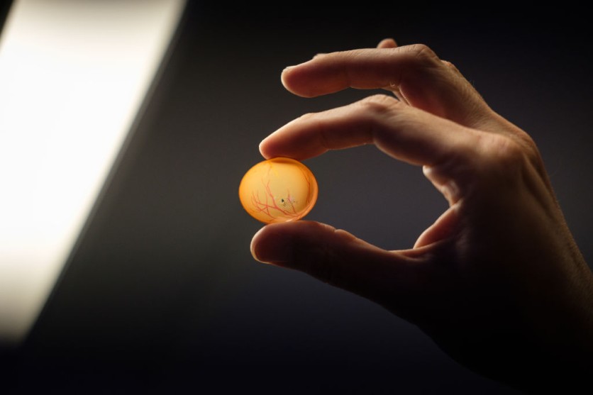
Scientists from the University of Wisconsin-Madison were able to reconnect lab-grown retina cells after separation, which may indicate that it is ready for human trials for those suffering from degenerative eye disorders, as per a press release.

Final Piece of the Puzzle
More than ten years ago, the research team discovered a method to create structured cell clusters, or organoids, that mimic the retina, the light-sensitive tissue in the back of the eye.
They coaxed layers of diverse retinal cell types to develop from human skin cells that had been altered to serve as stem cells. Retinal cells perceive light and ultimately transmit what we see to the brain.
The recent study is "the final piece of the puzzle," says David Gamm, the research's principal author.
Gamm said they wanted to use the cells from the organoids as replacement components for similar types of cells that have been lost due to retinal diseases.
In 2022, Gamm and UW-Madison collaborators published studies demonstrating that photoreceptors, retinal cells grown in a dish, respond to various light wavelengths and intensities similarly to healthy retinal cells.
They also showed that photoreceptors might communicate with new neighbors via characteristic biological cords known as axons after being separated from nearby cells in their organoids.
The retinal organoids were divided into individual cells, given a week to grow new connections and lengthen their axons, exposed to the virus, and were eventually examined.
The scientists discovered several retinal cells that were fluorescently colored, indicating that one had become infected with rabies across a synapse that had successfully developed between nearby cells.
Read also : Eye Disease Treatment and Prevention Through Smart LED Contact Lenses: How Effective Is It?
"The Next Clear Step"
"We've been quilting this story together in the lab, one piece at a time, to build confidence that we're headed in the right direction," Gamm said in a statement.
"It's all leading, ultimately, to human clinical trials, which are the clear next step."
The researchers confirmed the existence of synaptic connections and examined the cells involved.
Photoreceptors, or rods and cones, made up the bulk of the retinal cell types generating synapses; these cells are lost in disorders including retinitis pigmentosa, age-related macular degeneration, and other eye injuries.
Retinal ganglion cells, the second most prevalent cell type, deteriorate in diseases of the optic nerve such as glaucoma.
Gamm said that this was an important revelation for the team as it shows the potentially vast impacts that these retinal organoids pose.
The findings of the study are published in PNAS.
Related Article : Kuo's New Apple Leaks: Upcoming Mixed Reality Headset Could Have Advanced Eye Tracking System

ⓒ 2026 TECHTIMES.com All rights reserved. Do not reproduce without permission.


