UT Health Science Center San Antonio researchers have revealed an exceptional tool capable of counting brain lesions in a matter of seconds. This groundbreaking technology harnesses the power of artificial intelligence (AI) to precisely quantify and map brain lesions.
AI Tool Capable of Countine Brain Lesions within Seconds
Through this new AI tool, Interesting Engineering reported that the medical field will be able to continue offering valuable information about conditions like cerebral small-vessel disease, linked to the development of stroke and dementia, but now more efficiently.
UT Health Science Center's Glenn Biggs Institute for Alzheimer's and Neurodegenerative Diseases Professor Dr. Mohamad Habes stated, "Certain kinds of brain lesions are tremendously difficult to quantify without AI." As an assistant professor of radiology, he contributed a lot to this research.
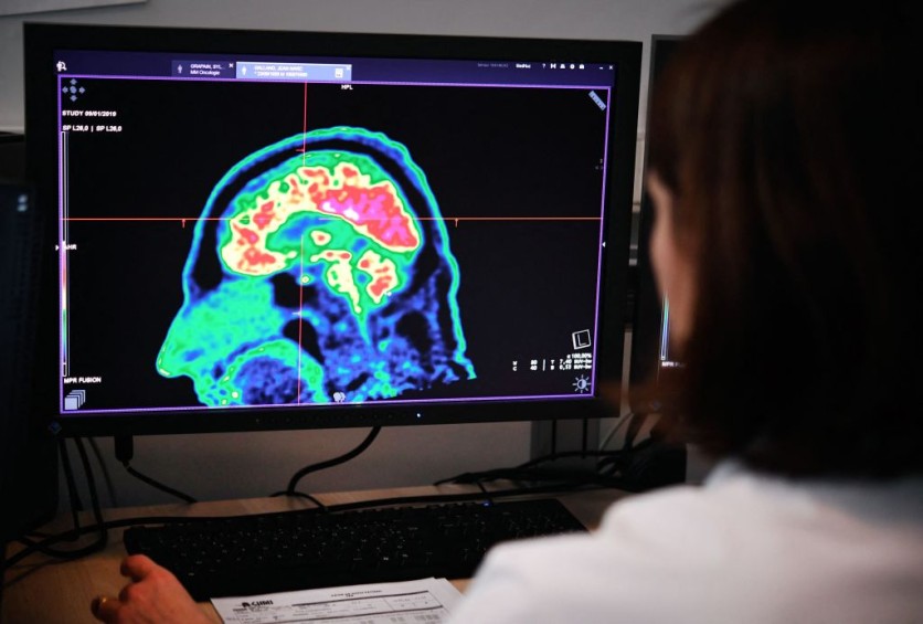
The research demonstrated how an AI tool can be valuable in detecting and quantifying enlarged perivascular spaces (ePVSs). These spaces, which contain cerebrospinal fluid, enclose blood vessels and act as indicators for cerebral small-vessel disease.
The AI tool used in the study likely employed machine learning algorithms to process and analyze brain imaging data. By training the AI model on a large dataset of brain images, it could learn to identify and count ePVSs accurately.
As per the UT Health San Antonio website, this involved 1,026 participants who were part of the Multi-Ethnic Study of Atherosclerosis (MESA). The study collaborated with researchers from eight different institutions, indicating a collaborative effort to investigate the utility of AI in analyzing ePVSs.
The researchers have expressed their aspirations to conduct additional studies on the AI tool used to count brain lesions at the Alzheimer's Disease Research Centers (ADRCs). These centers, designated as Centers of Excellence by the U.S. National Institute on Aging, aim to further investigate the potential of the AI tool, including the South Texas ADRC.
Emergence of AI in Medicine
AI has been making significant strides in medical imaging analysis, including the interpretation and diagnosis of various conditions. One area where AI has shown promise is in the detection and quantification of brain lesions, which can be indicative of different neurological disorders or injuries.
Traditionally, the process of counting brain lesions could be time-consuming and require the expertise of radiologists or other specialists. AI tools can help automate and expedite this process by leveraging advanced algorithms and machine learning techniques.
These tools are trained on large datasets of brain images, enabling them to recognize and classify different types of lesions accurately. Through AI, this could potentially streamline the diagnosis and treatment planning process, allowing healthcare professionals to make informed decisions more efficiently.
However, it's important to note that the development and implementation of AI tools in the medical field must undergo rigorous testing, validation, and regulatory approval processes to ensure their safety, reliability, and efficacy. This study was published in JAMA Network Open.

ⓒ 2026 TECHTIMES.com All rights reserved. Do not reproduce without permission.


