Researchers have introduced a novel technology capable of implanting minute "marks" in regions of the brain where activity has been recorded, presenting a breakthrough in neuroscience.
According to Medical Xpress, the innovative method involves passing a microcurrent through tungsten electrodes inserted into the brain to record neural activity.
Unlike existing techniques, these marks do not damage brain tissue and can be placed safely inside the brain. This technology facilitates high-resolution mapping of the distribution of neurons with specific functional characteristics.
'Marks' in the Brain
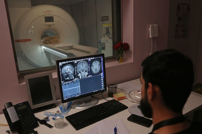
Studies monitoring deep brain regions, such as the hippocampus and thalamus, traditionally utilize needle-like microelectrodes for precise recordings.
While providing higher spatiotemporal resolution than alternatives like fMRI or EEG, these electrodes face challenges in determining their exact location post-removal.
To address this, the common practice of "marking" is used, but it suffers from limited spatial resolution and can damage brain tissue, restricting spatial precision to approximately 0.1 mm.
The breakthrough comes from the collaboration of researchers Tatsuya Oikawa, Toshimitsu Hara, Kento Nomura, and Associate Professor Kowa Koida from the Department of Computer Science and Engineering at Toyohashi University of Technology.
They focused on the electrolysis of tungsten, a material commonly used for needle-like electrodes. The method involved using electrolysis to deposit oxidized tungsten within deep brain tissues.
In the process of electrolysis, the tungsten electrode produces metal oxide and hydrogen bubbles during positive and negative currents, respectively. The alternating application of these currents leads to the creation of a small oxide lump at the tip of the electrode, serving as a marker for accurately identifying its specific location.
In experiments conducted on the brains of mice and monkeys, the scientists effectively implanted tiny oxide lumps with diameters as small as 20 µm without causing harm to brain tissue.
Notably, they observed that these oxide deposits displayed a red luminescence when subjected to a standard brain tissue stain. This unique feature makes them easily discernible from the surrounding background when observed under low-power microscopy.
Read Also : [STUDY] Food Packaging Plastic Entering Brain Could Increase Neurodegeneration Risks, Claim Experts!
Accidental Discovery
Kowa Koida, the team's advisor, highlighted the accidental discovery during experimentation, saying, "Despite this setback, we observed the formation of a mark.
Upon closer consideration, we recognized that electrolysis, which is commonly used to form tungsten rods into thin needles for electrodes, produces an oxide powder."
The researchers are now applying this technology to explore the microscopic functional structure of the lateral geniculate nucleus in macaque monkeys, aiming to demonstrate its efficacy for widespread adoption in neuroscience.
The research team further noted that the technique's safety has already been established, and its combination of measurement and deposition processes may find applications in medical care, leveraging in vivo metal electrolysis for various procedures, such as treating aneurysms.
The team's findings were published in the journal eNeuro.
Related Article : Astronauts' Brains Change During Space Missions? New Study Claims Cosmonauts Already Experience Brain Rewiring

ⓒ 2026 TECHTIMES.com All rights reserved. Do not reproduce without permission.


