Is it an eye? Or is it a painting that deserves to be hung at the Louvre? Well, get ready to be amazed by the stunning image of the human retina that looks like an absolute work of art!
Researchers from ETH Zurich and the Universities of Zurich and Basel have created a new method to collect and organize information about organoids, which are small three-dimensional tissues that resemble human organs. They used this method to study the organoids of the human retina.
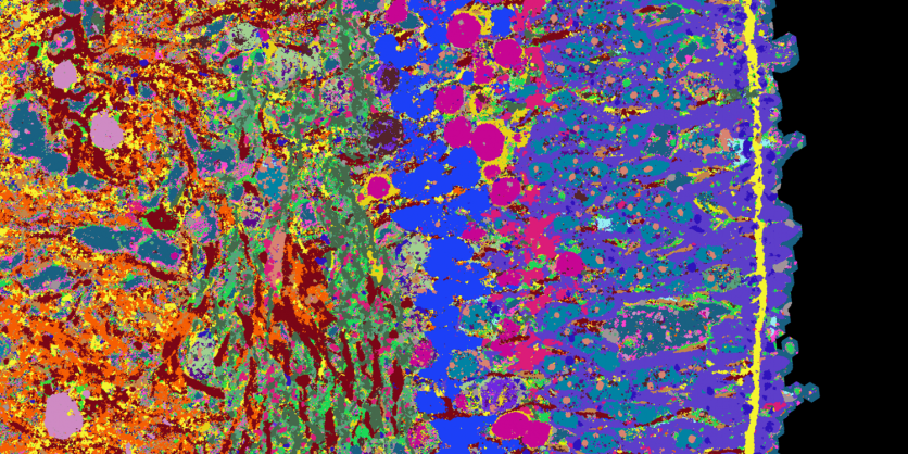
4i Technology
The scientists used a technique called 4i technology to see many proteins in a small piece of tissue at a high level of detail using fluorescence microscopy. This technique helped them see and study 53 different proteins in a tissue sample, which showed them how the cells in the retina, like rods, cones, and ganglion cells, work.
"This new imaging technique can visualize several dozen proteins in a thin tissue section at high resolution using fluorescence microscopy," said Barbara Treutlein, Professor of Quantitative Developmental Biology at the Department of Biosystems Science and Engineering at ETH Zurich in Basel.
"We can use this time series to show how the organoid tissue slowly builds up, where which cell types proliferate and when, and where the synapses are located. The processes are comparable to those of retinal formation during embryonic development," said Gray Camp, a professor at the University of Basel and a senior author of the study.
Professor Treutlein explained that the researchers created a series of images and genetic information that shows how retinal organoids develop over 39 weeks. They can change their growth and test drugs on them to learn about healthy tissue and diseases.
The researchers aim to use this approach on other tissues, like different parts of the brain and various tumor tissues, in the future.
Read Also : Researchers Use Machine Learning to Detect Flaws in Laser Powder Bed Fusion Additive Manufacturing Process
Healthy Retina
Currently, the team is examining the development of a healthy retina. However, in the future, they plan to disrupt the development of retinal organoids using drugs or genetic modifications.
This will allow them to gain a better understanding of diseases like retinitis pigmentosa, which causes the light-sensitive receptors in the retina to deteriorate, leading to blindness. The team aims to determine when this process begins and how it can be halted.
The comprehensive map of human organoids and tissues is expected to give crucial knowledge on how human tissue types develop and provide an understanding of the cause and progression of diseases, leading to potential treatments.
The study titled "Multimodal spatiotemporal phenotyping of human retinal organoid development" was published in the journal Nature Biotechnology.
Related Article : The First 'Vagina-on-a-Chip' May Aid Researchers in Testing Drugs for Bacterial Vaginosis

ⓒ 2026 TECHTIMES.com All rights reserved. Do not reproduce without permission.


